Translate this page into:
Case of shoulder pain
Corresponding Author:
Nizar A Al-Nakshabandi
Department of Radiology, College of Medicine, King Saud University, Riyadh
Saudi Arabia
nizar97@hotmail.com
| How to cite this article: Al-Nakshabandi NA. Case of shoulder pain. J Musculoskelet Surg Res 2020;4:61-62 |
History
A 29-year-old male complained of shoulder pain. He has no history of recent trauma. Pain is gradually increasing with activity.
He is known to have sickle cell disease.
- What are your findings?
- What is the differential diagnosis?
- What are the causes?
Findings
Plain radiograph of the left shoulder [Figure - 1]a demonstrates periarticular osteopenia. In addition, subchondral lucency is seen in the humeral head (a crescent sign of avascular necrosis [AVN]).
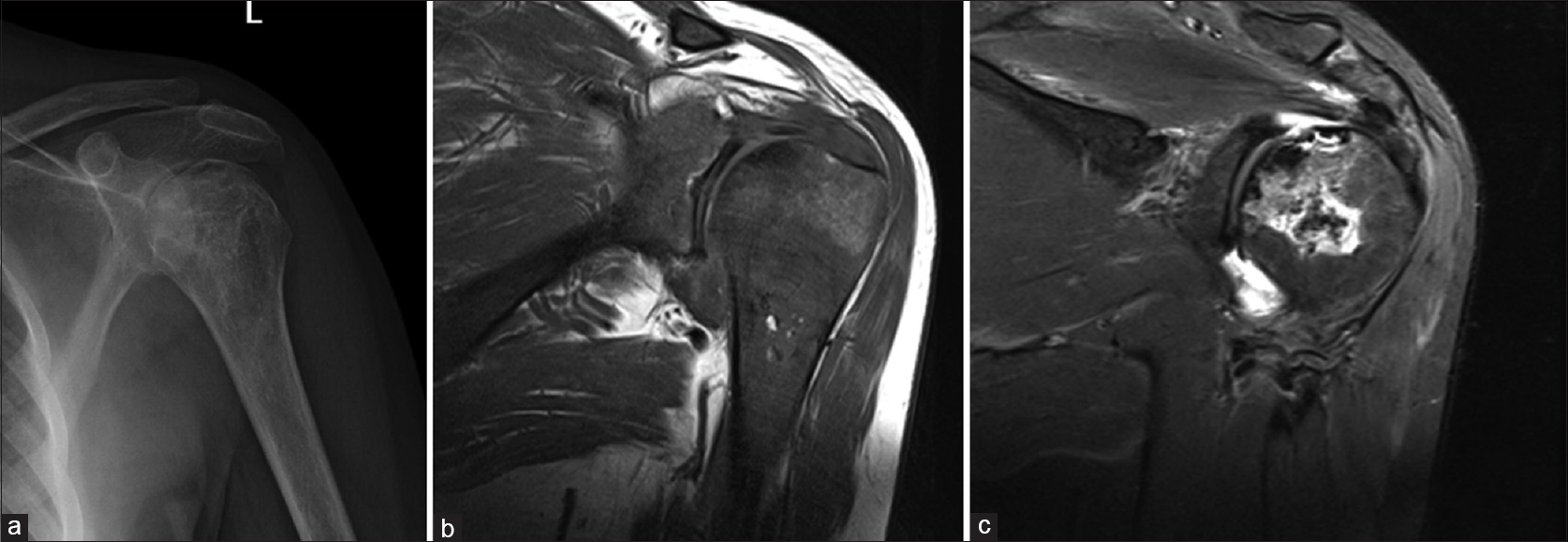 |
| Figure 1 |
Coronal T1-weighted [Figure - 1]b image shows diffuse low signal changes involving the whole left shoulder consistent with edema.
Coronal T2-weighted [Figure - 1]c image shows a mixture of low and high signal changes in the humeral head representing the double line sign, which is the serpiginous outer dark signal and bright inner signal (granulation tissue). In addition, the crescent sign that was seen on the plain radiograph becomes the rim sign on T2WI, which is the result of osteochondral fragmentation.
Differential Diagnosis
- AVN
- Infection (osteomyelitis with or without septic arthritis)
- Neoplasm - less likely.
Diagnosis
The patient underwent Stage III b AVN of the left shoulder.
Causes of Avascular Necrosis
There are many mnemonics; I discourage you from using them unless you are stuck. The reason is you may mention the least cause first in your region, such as Caisson disease (divers' disease) in a desert country, which will make you look strange in front of the examiner. The way I remember it is by imaging a blood vessel, so in the vessel is red blood cell if defected then – sickle cell disease, medications – exogenous steroids, indigenous steroids (Cushing's disease), and finally in the blood alcohol can run. Vessel wall – vasculitis. Outside vessel – trauma, and radiotherapy.
There are other causes such as pregnancy-related AVN, pancreatitis, Gout, and Gaucher's disease, but if you mention them before mentioning sickle cell disease, the examiner will not be pleased.
Discussion and Pearls
Remember that examiners like testing a common disease in an uncommon location. Hence be aware that AVN usually affecting the hips can also involve the shoulders.
In cases of nontraumatic AVN, bilateral involvement is common.
Always screen the other side on magnetic resonance imaging.
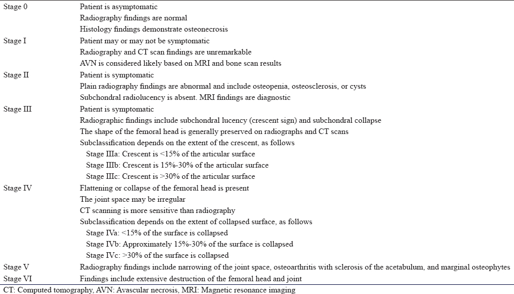
Many staging systems used, the most comprehensive is the Association Research Circulation Osseous staging system is used to grade AVN.
Declaration of patient consent
The author certifies that he has obtained all appropriate patient consent forms. In the form, the patient has given his consent for his images and other clinical information to be reported in the journal. The patient understands that his name and initials will not be published and due efforts will be made to conceal his identity, but anonymity cannot be guaranteed.
Financial support and sponsorship
Nil.
Conflicts of interest
There are no conflicts of interest.
Further Reading
Further Reading
- Bonakdarpour A, Reinus WR. Diagnostic Imaging of Musculoskeletal Diseases: A Systematic Approach. Springer; 2010.
- Resnick D, Kransdorf MJ. Bone and joint imaging. WB Saunders Co.; 2005.
- Kaplan P. Musculoskeletal MRI. WB Saunders Co.; 2001.
Fulltext Views
965
PDF downloads
350


![[Figure - 1]](#fig_SaudiOrthopJ_2020_4_1_61_278385_f1.jpg)